
How Is a Jaw Cyst Treated? The Surgical Process

Summary
General information on the overall approach to treating jaw cysts, the surgical methods of enucleation and marsupialization, why the removed tissue is sent for pathology, and the healing process.
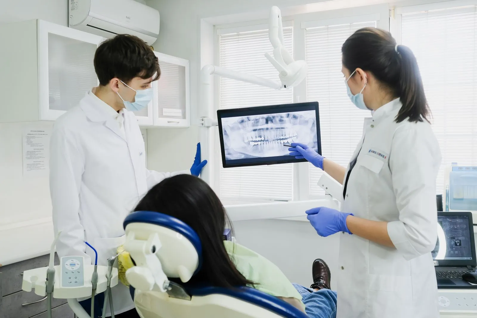
Treating a jaw cyst is a process that aims to remove the sac-like formation developing within the jawbone or around the roots of teeth using appropriate surgical methods, or, in the case of large cysts, to first reduce it by emptying it. The method to be used is determined after examination and radiographic evaluation, according to the type, size and location of the cyst and its relationship with neighboring anatomical structures.
As part of his work in periodontology and oral surgery, Uzm. Dt. Emre Karakaya focuses on the evaluation of jaw cysts and their surgical treatment. The headings below are intended for general information; they are not a substitute for a personal examination, and consulting your dentist for accurate information is recommended.
The General Approach to Treating Jaw Cysts
The general approach in treating jaw cysts is to remove the cyst surgically while protecting, as far as possible, the bone tissue in the area where it is located. In small, well-defined cysts the formation can often be removed as a whole in a single session, whereas in large cysts or those adjacent to important anatomical structures a staged approach may be preferred. The treatment decision is made by taking into account the cyst's diagnosis, size and relationship with the surrounding tissues.
Because the type and mechanism of the cyst directly affect the treatment plan, a detailed evaluation is carried out before surgery; the general characteristics of cysts and how they are noticed are covered in the article on what a jaw cyst is. The procedure is most often planned under local anesthesia, and its scope and duration vary from person to person.
Surgical Methods: Enucleation and Marsupialization
Enucleation describes a method in which the membrane surrounding the cyst (the cyst sac) is carefully separated from the surrounding bone and removed as a whole. This approach is often considered in cysts with clear boundaries that can be separated from the surrounding tissue; the cavity that forms in the area may fill in over time as the bone heals. During the procedure, care is taken to protect neighboring structures such as tooth roots, the nerve canal and the sinus cavity.
Marsupialization, on the other hand, is based on opening part of the cyst to drain its contents and reduce the internal pressure, particularly in large cysts or formations located close to important anatomical structures; in this way the cyst may shrink over time and, if needed, be removed at a second stage. Which method is suitable and how the jaw cyst surgery process is planned are determined individually, according to the examination and imaging findings.
Why Pathological Examination Is Done
The tissue removed during surgery is routinely sent for pathological examination. This examination makes it possible to determine the type of the formation with certainty at a microscopic level and to assess whether it is benign. Although radiographic images provide information about the boundaries of the cyst, a definitive diagnosis is clarified through the histopathological evaluation of the tissue sample.
The result of the pathological examination helps shape the follow-up plan. While some cyst types may require closer monitoring, the results and the recommended check-up intervals vary from person to person. This evaluation is regarded as an important stage for the completeness of the treatment process.
Healing and Follow-up
After the surgical procedure, some swelling, tenderness or limited discomfort may be seen in the area; these findings are usually a natural part of the healing process. Following the oral hygiene practices and care instructions recommended by the dentist supports healing. The healing time varies according to the size of the cyst, the method applied and the person's general state of health.
Bone healing in the treated area is monitored over time with radiographic check-ups, and the frequency of follow-up is planned according to the case. Results vary from person to person; the information here is intended for general information and is not a substitute for examination. Consult your dentist for accurate information and a treatment plan suitable for you.
This content is for informational purposes only and does not replace an examination by a dentist. The process and results vary from person to person.
Reading this while planning treatment in Turkey? See the Denizli visit planner and the dental tourism guides.
Related Treatments
Write to us with your questions
You can get in touch via WhatsApp, phone or email for information about treatments and the process.