Skip to content
Uzm. Dt. Emre Karakaya — Periodontoloji & İmplant Cerrahisi

Jaw Cyst Surgery

Surgical treatment and follow-up of jaw cysts.

Jaw Cyst Surgery

Jaw cyst surgery involves diagnosing cysts that can develop within the jawbone and around the roots of teeth, and removing them surgically. Cysts are sac-like formations filled with fluid or soft tissue, and they often cause no symptoms, being noticed during radiographic examinations.

As part of his work in periodontology and oral surgery, Uzm. Dt. Emre Karakaya deals with the assessment and surgical treatment of jaw cysts. The aim is to determine the type and boundaries of the cyst radiologically and then remove it with a suitable surgical method while taking care of the surrounding tissues. This content is for information only; consult your physician for a definitive assessment.

What Is a Jaw Cyst and What Are Its Types?

A jaw cyst is a cavity that forms within the jawbone or around the roots of teeth, surrounded by a membrane (epithelium) and filled with fluid or a semi-solid material. It can grow over time and thin the bone in the area where it is located. Most cysts are benign; however, depending on their size and location, they can affect neighboring teeth, nerves and bone structures.

Different types of cysts can be seen in the mouth. A radicular (periapical) cyst usually develops around the root tip of a tooth that has lost its vitality due to decay or trauma, in connection with an inflammatory process. A dentigerous (follicular) cyst can form around the crown of an unerupted or impacted tooth. Because identifying the type affects the treatment plan, a definitive diagnosis is made through radiographic and pathological evaluation.

Radiographic Diagnosis

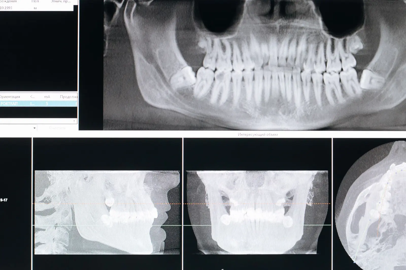
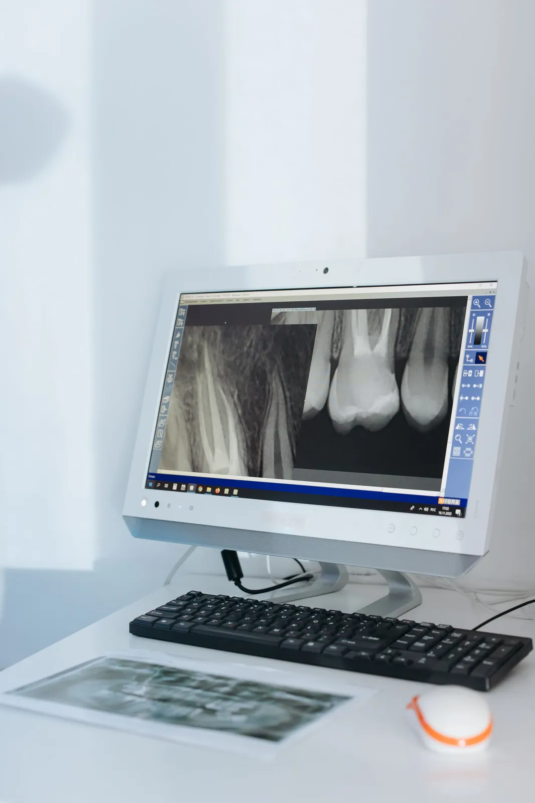
Because jaw cysts often progress silently, they are frequently noticed incidentally on X-rays taken for another reason. A panoramic X-ray is the first step in assessing the location and general boundaries of the cyst. When needed, the relationship between the root and the cyst is examined in more detail with periapical films.

When clearer information is needed about the size of the cyst, its relationship with neighboring anatomical structures (nerve canal, sinus cavity, tooth roots) and its three-dimensional boundaries, cone-beam computed tomography (CBCT) may be used. Radiographic evaluation forms the basis of surgical planning; however, the type of the cyst is confirmed only by the pathological examination of the removed tissue.

Surgical Methods: Enucleation and Marsupialization

There are two main approaches in the surgical treatment of jaw cysts. Enucleation is the removal of the cyst sac as a whole by separating it completely from the surrounding tissue, and it is a method considered for many cysts. The bone cavity that forms afterwards is left to the healing process over time.

Marsupialization is a method that may be considered particularly for large cysts or those adjacent to important anatomical structures. In this approach, the cyst cavity is opened into the mouth to reduce the internal pressure and allow the cyst to shrink over time; complete removal may be planned in a second stage when needed. Which method is suitable depends on the type, size and location of the cyst and is determined by examination.

Treatment Stages

1) Radiographic Evaluation: First, the location, size and relationship of the cyst with neighboring structures are examined with a panoramic X-ray and, when needed, tomography. This stage provides a preliminary assessment of the boundaries and type of the cyst.

2) Surgical Planning: In light of the radiographic findings, the surgical method to be applied (enucleation or marsupialization), the choice of anesthesia and any additional procedures are planned. The condition of the related teeth and the general medical history are evaluated at this stage.

3) Local Anesthesia: The treatment area is numbed with local anesthesia, so that no pain is felt during the operation. The anesthesia method is determined according to the person's condition.

4) Cyst Operation: Depending on the planned method, the cyst sac is removed or emptied and reduced in size. During the procedure, care is taken of the surrounding bone, tooth roots and nerve structures; additional treatments for the relevant tooth are evaluated when needed.

5) Pathology and Follow-up: The removed tissue is sent for pathological examination. Follow-up appointments are planned at certain intervals to monitor healing and assess bone repair.

Pathological Examination and Follow-up

The cyst tissue removed surgically is routinely sent for pathological examination. This examination makes it possible to determine the type of the formation with certainty and to assess whether it is benign. Although the radiographic appearance gives a preliminary idea, a definitive diagnosis is made only through microscopic examination of the tissue.

During recovery after the operation, the repair of the bone cavity and the condition of the area are monitored with check-ups at certain intervals. Because some types of cysts carry a possibility of recurrence, regular check-ups are an important part of the process. The frequency and duration of follow-up vary from person to person; consult your physician for accurate information.

Frequently Asked Questions

A jaw cyst is a sac-like formation that develops within the jawbone or around the roots of teeth, surrounded by a membrane and filled with fluid or soft tissue. Most are benign, but they can grow over time and affect the surrounding bone and teeth. The type and treatment are determined by examination.

Jaw cysts often cause no symptoms and are frequently noticed on an X-ray taken for another reason. As a cyst grows, swelling, a feeling of fullness in the jaw, a change in tooth position, or discomfort due to pressure may occur. Symptoms vary with the type and size of the cyst.

Untreated cysts can grow over time and thin the surrounding bone, and may affect neighboring teeth and nerve structures. For this reason, it is advisable to have a detected cyst evaluated by a clinician. How the process progresses depends on the type of cyst; consult your physician for accurate information.

Because the procedure is performed under local anesthesia, the aim is that no pain is felt during the operation. Mild swelling or discomfort may occur afterwards, and this is managed with the care recommended by your clinician. The experience varies from person to person.

In the first days, mild swelling or tenderness may be seen in the area, and this usually eases within a few days. Repair of the bone cavity is a longer process that varies with the size of the cyst. Follow-up appointments are planned to monitor healing; the duration varies from person to person.

Pathological examination of the removed cyst tissue is performed to determine the type of the formation with certainty and to assess whether it is benign. Although the radiographic image provides preliminary information, a definitive diagnosis is made by microscopic examination. This examination also helps in creating an appropriate follow-up plan.

This content is for informational purposes only and does not replace a physician examination. The process and results vary from person to person.

Share

Related Posts

View All
What Is a Jaw Cyst?

What Is a Jaw Cyst?

General information on what jaw cysts are, their common types, how they often develop without symptoms, and how they are noticed on radiographs.

Read More