
Panoramic X-Rays and Dental Tomography: When Are They Considered?

Summary
What do panoramic X-rays and cone-beam computed tomography (CBCT) show, and in which situations are they considered? General information on their scope, differences and radiation dose.
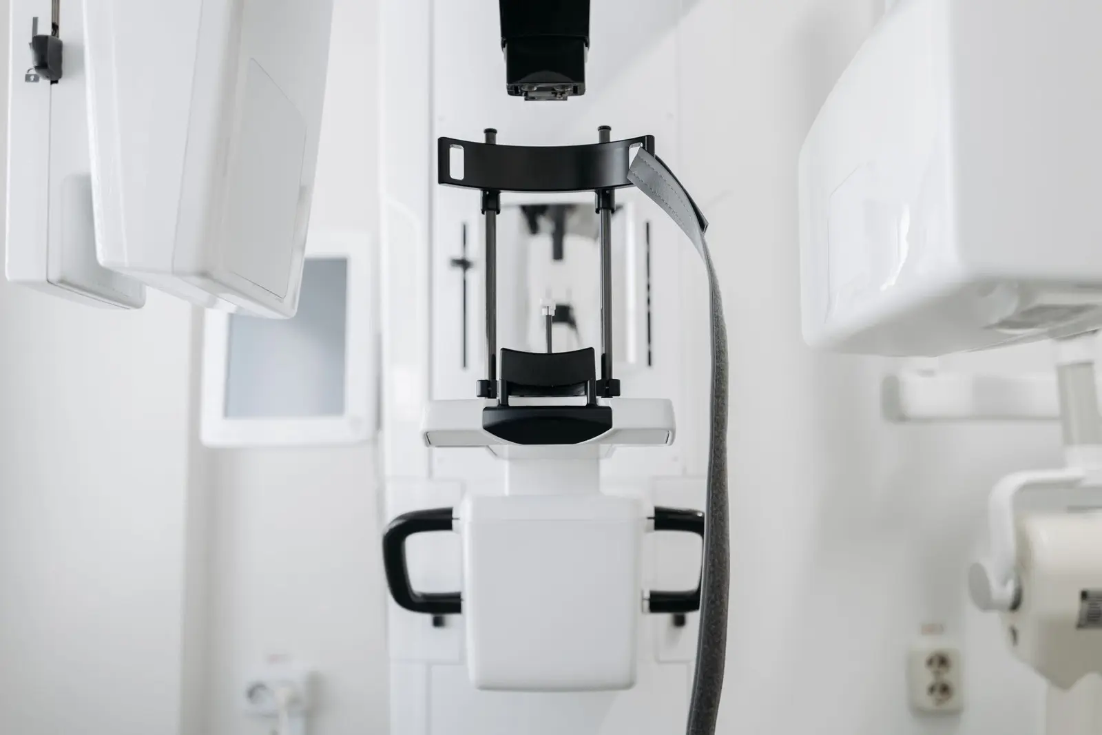
During a dental examination, structures that cannot be seen directly — tooth roots, the jawbone, impacted teeth and the sinus regions — are assessed with the help of radiographic imaging. While the small films placed inside the mouth show individual teeth in detail, panoramic X-rays and dental cone-beam computed tomography (CBCT), taken from outside the mouth, allow the jaws to be assessed within a wider frame.
This article outlines what each method shows, in which situations they are considered, the differences between them and the general approach to radiation dose. The content is for information purposes; whether any imaging is necessary is assessed by the dentist together with the clinical examination findings.
What Does a Panoramic X-Ray Show?
A panoramic X-ray (orthopantomogram) shows the upper and lower jaws, all the teeth, the jaw joints and the sinus floors together in a single two-dimensional image. During the exposure the device rotates around the head and lays the curved arch of the jaws out on one image, providing an overall view of the mouth.
It is frequently used in routine assessment, in observing the presence and general position of impacted teeth, in screening for decay and bone loss, and as a first step in planning extractions or treatment. Because it is a two-dimensional image, however, structures may overlap; depth information — how close a lesion lies to the cheek or tongue side — cannot be read from it directly.
What Is Dental Cone-Beam Tomography (CBCT)?
Dental volumetric tomography, also known as cone-beam computed tomography (CBCT), produces a three-dimensional image of the area of interest. The data can be examined as cross-sections on different planes; the thickness and height of the bone can be measured in millimetres, and the position of anatomical structures relative to one another can be followed in three dimensions.
It is considered when a question requires three-dimensional detail, such as the course of the nerve canal, the relationship between the sinus floor and tooth roots, or the contact of an impacted wisdom tooth with neighbouring structures. The field of view can be kept narrow or planned to cover a wider region, according to need. The exposure itself is usually brief; keeping still during it matters for a clear image.
Which One Comes Up in Which Situation?
Rather than being alternatives to each other, the two methods answer different questions; each has its own scope and limits. The panoramic X-ray offers a general overview, while CBCT focuses on the three-dimensional detail of a specific area. The process therefore often starts with a panoramic image; if three-dimensional information is needed after this image and the examination are assessed together, tomography is planned separately.
Implant surgery planning is a typical example: tomography may be used when the volume and density of the bone and its relationship with neighbouring anatomical structures such as the nerve canal or sinus floor need to be assessed in three dimensions. Similarly, defining the borders of a cyst within the jaw, cyst follow-up and the assessment before cyst surgery are among the situations in which tomography may be considered.
Radiation Dose and the ALARA Principle
The imaging methods used in dentistry generally fall within the low-dose range of medical imaging; the dose also varies with the device, the size of the imaging field and the settings chosen. Even so, the fact that every radiographic examination involves ionising radiation is not disregarded; imaging is therefore planned when there is a clinical question the examination findings cannot answer.
This approach is summarised by the ALARA principle — as low as reasonably achievable: the imaging field and the dose are kept at the lowest level that will answer the clinical question. Which method is used, when and over which area is assessed by the dentist by weighing the benefit of the information to be obtained. Under the same principle, if a suitable recent image already exists, using it instead of taking a new one may also be considered. Informing the dentist of pregnancy, or the possibility of pregnancy, before the exposure is also part of this assessment.
The Importance of Incidental Findings
Radiographic images may also reveal findings beyond the reason they were taken. Impacted teeth that have gone unnoticed, inflammatory changes at the root tip or cystic lesions that can grow quietly within the jaw may appear incidentally on a panoramic image taken for another purpose.
Some of these lesions can progress for a long time without causing complaints; this subject is discussed in detail in the article on jaw cysts without symptoms. When an incidental finding is detected, whether it calls for follow-up or further assessment is determined by the dentist according to its nature and size.
In Summary
Panoramic X-rays and dental cone-beam tomography are two tools of the same diagnostic process that answer different questions. Which one is needed, and when, varies from person to person depending on the clinical findings, the planned procedure and the anatomy of the area.
This content has been prepared for general information purposes and does not replace an examination; consult your dentist for accurate information on the imaging approach suitable for you.
This content is for informational purposes only and does not replace an examination by a dentist. The process and results vary from person to person.
Reading this while planning treatment in Turkey? See the Denizli visit planner and the dental tourism guides.
Write to us with your questions
You can get in touch via WhatsApp, phone or email for information about treatments and the process.