
When Should Wisdom Teeth Be Removed and When Can They Be Monitored?

Summary
There is no single rule on whether an impacted wisdom tooth must be removed: symptom-free teeth with no concerning relationship to neighboring structures can be monitored with regular check-ups, while findings such as recurrent pericoronitis, decay in the neighboring molar or cystic enlargement of the follicle support extraction. The decision is made individually by weighing the tooth's position, complaints and radiographic findings together; consult your dentist.
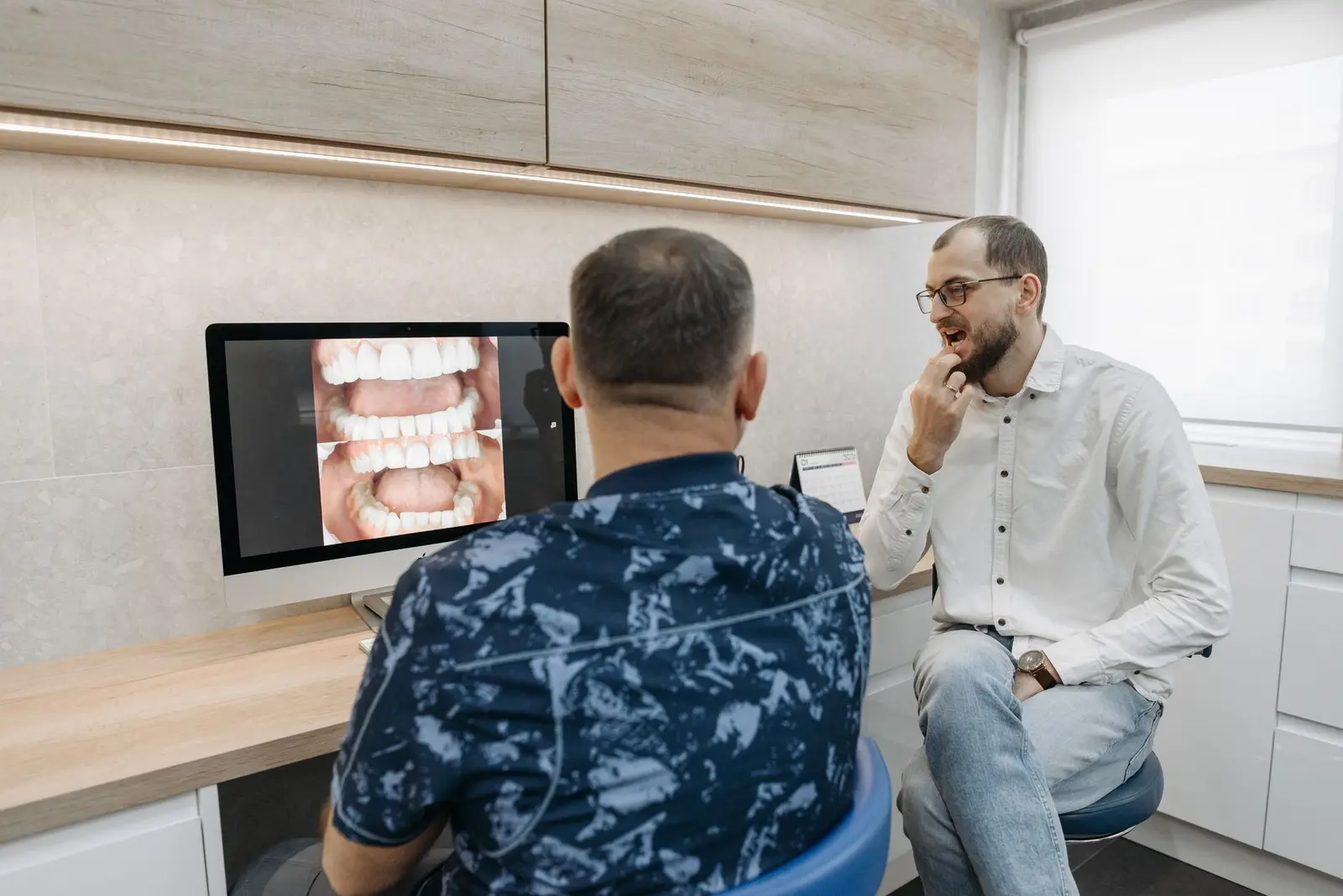
One of the questions asked most often about impacted wisdom teeth is whether the tooth always has to be removed. There is no single answer: some impacted teeth can be monitored for years with regular check-ups, while in others the findings point toward extraction. The decision is made individually, weighing the tooth's position, the person's complaints and the radiographic findings together.
This article focuses on the choice between monitoring and extraction; why wisdom teeth remain impacted and how the surgical process works are covered in the article on impacted wisdom teeth. The headings below are for general information; the right approach for you can only be determined through clinical examination and imaging.
Can a Trouble-free Impacted Tooth Be Monitored?
Some impacted teeth that cause no symptoms, remain completely within the bone and show no concerning relationship with neighboring structures can be followed with regular check-ups instead of being removed, avoiding surgery for a tooth considered unlikely to cause problems. Monitoring, however, does not happen by default: it is a conscious clinical decision made after examination and imaging.
Nor is monitoring passive waiting. Because the tooth's position and its relationship with surrounding tissues can change over time, periodic examinations and radiographic checks when needed are recommended. In partly erupted teeth connected to the gum, contact with the oral environment makes bacterial accumulation more likely, so the decision to monitor calls for extra care.
Findings That Support Extraction
Recurrent episodes of pericoronitis — repeated inflammation of the gum around a partly erupted tooth — are among the leading findings that support extraction. Even when each episode settles, the underlying cause remains as long as the tooth's position is unchanged, and the inflammation can recur over time.
In addition, decay in the neighboring second molar related to an area that cannot be cleaned, and root-surface changes associated with long-standing pressure can also develop. Cystic enlargement of the follicle tissue around the tooth is another radiographic finding assessed in favor of extraction. None of these findings is interpreted alone; they are weighed together with the clinical examination.
The Decision and Imaging Process
The first step is usually a panoramic X-ray, on which the tooth's position, its relationship with neighboring teeth and the surrounding bone are assessed in general terms. In the lower jaw, when the tooth appears close to the nerve canal, cone-beam computed tomography (CBCT) may be used to view this relationship in three dimensions.
When the findings favor removal, impacted wisdom tooth extraction is a planned surgical procedure usually performed under local anesthesia; the points recommended for the period afterward are described in the article on healing after extraction. Whether monitoring or extraction is appropriate varies from person to person; consult your dentist about your own situation.
Questions About This Article
There is no standard interval; monitoring is not passive waiting. Because the tooth's position and its relationship with surrounding tissues can change over time, periodic examinations and radiographic checks when needed are recommended. The follow-up interval is determined by your dentist according to the tooth's condition.
The settling of an episode does not mean the problem is resolved; as long as the tooth's position is unchanged, the inflammation can recur over time. Recurrent pericoronitis is among the findings that support extraction; the frequency and severity of episodes are weighed, and the decision is evaluated together with your dentist.
Not in every case. The first step is usually a panoramic X-ray; in the lower jaw, when the tooth appears close to the nerve canal, cone-beam computed tomography may be used to view this relationship in three dimensions. The need for imaging is determined by the examination findings.
This content is for informational purposes only and does not replace an examination by a dentist. The process and results vary from person to person.
Reading this while planning treatment in Turkey? See the Denizli visit planner and the dental tourism guides.
Write to us with your questions
You can get in touch via WhatsApp, phone or email for information about treatments and the process.